Mosquito Anatomy
Before we jump into detailed anatomy, let's look at five main characteristics of mosquitoes:
- They have a hard outer shell called an exoskeleton.
- They have three main body parts: head, thorax, abdomen.
- They have a pair of antennae that are attached to their head.
- They have three pairs of legs used for walking.
- They have one pair of wings used for flying.
You can use the illustrations below to explore the anatomy of the mosquito, both what you can see from the outside and also the parts of the mosquito located inside.
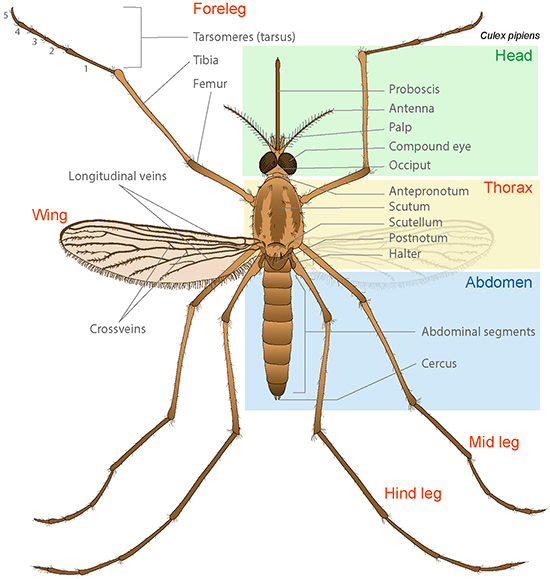
Labeled illustration of the external anatomy of a mosquito. Image modified from LadyofHats via Wikimedia Commons.
Looking at the Outside of an Adult Mosquito
| Head | Location of the eyes and brain, and where the antennae attach. | ||
| Proboscis | Tube-like mouth part used to suck up fluids. In females, this is strong enough that it can pierce skin. | ||
| Antenna | Movable segmented feather-like feeler that detects chemicals like carbon dioxide and air currents. | ||
| Palp | A sensory organ that can detect odors and can feel objects. | ||
| Compound Eye | Eyes made of many light detectors called ommatidia. | ||
| Occiput | The back of the head. | ||
| Thorax | Midsection where the (6) legs and (2) wings attach. | ||
| Antepronotum | A protective plate that sits on top of the front of the thorax. | ||
| Scutum | The large front section of the thorax. | ||
| Scutellum | The middle section of the thorax. | ||
| Postnotum | The end of the thorax, which also holds the septum that separates the thorax and the abdomen. | ||
| Halter | An organ that helps mosquitoes to steer while they fly. | ||
| Abdomen | The hind part of the mosquito, where most of digestion, eliminating waste, and reproduction occurs. | ||
| Abdominal segments | Sections of the abdomen. Each segment has a top and a bottom that are separated, allowing the abdomen to expand with a meal or with eggs. | ||
| Cercus | A projection from the end of the abdomen that is involved in mating and in egg-laying. | ||
| Tarsomeres | Moveable parts of the end of the leg. | ||
| Tarsus | The last segment of the leg and what touches the walking surface. | ||
| Tibia | Fourth segment of an insect leg; the tibia of the hind leg holds the pollen basket, where pollen is carried. | ||
| Femur | Third segment of an insect leg. | ||
| Foreleg | Leg located closest to the head. | ||
| Mid Leg | Leg located between the foreleg and hind leg. | ||
| Hind Leg | Leg farthest from the head. | ||
| Wing | Flat projections from the body that are used in flying. | ||
| Longitudinal Veins | Thickenings in the wing that lie along the long axis of the wing. | ||
| Crossveins | Thickenings in the wing that lie along the short axis of the wing. | ||
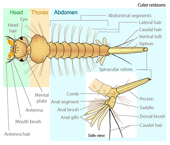
Labeled illustration of the external anatomy of a mosquito larva. Image modified from LadyofHats via Wikimedia Commons.
Looking at the Outside of a Mosquito Larva
| Head | Location of the eyes and brain, and where the antennae attach. | ||
| Eye | Larvae have fairly simple eyes called ocelli that sense light. | ||
| Head hair | Bits of hair near the front of the face. Hairs are used for sensing. | ||
| Mental plate | A protective plate on the top of the head. | ||
| Antenna | Movable segmented feather-like feeler that detects chemicals like carbon dioxide and air currents. | ||
| Mouth brush | A collection of hairs around the mouth that help with moving water and filter feeding. | ||
| Antenna hair | Hair on the antennae that help with sensing under water. | ||
| Thorax | Midsection where the (6) legs and (2) wings will later attach. | ||
| Abdomen | The hind part of the mosquito, where most of digestion, eliminating waste, and reproduction occurs. | ||
| Abdominal segments | Sections of the abdomen. Each segment has a top and a bottom that are separated, allowing the abdomen to expand with a meal. | ||
| Lateral hair | Longer bits of hair on the sides of the thorax and abdomen. Hairs are used for sensing. | ||
| Caudal hair | Paired long hairs near the end of the abdomen. Hairs are used for sensing. | ||
| Ventral tuft | Paired thin hair tufts near the end of the abdomen. Hairs are used for sensing. | ||
| Siphon | A breathing tube that larval mosquitoes use; it uses water tension at the surface to attach there. | ||
| Spiracular valves | Valves that can close off the end of the siphon, so it does not take on water. | ||
| Comb | A patch of small spine-like scales near the end of the abdomen. | ||
| Anal segment | The last segment on the abdomen. | ||
| Anal brush | A collection of hairs at the rear of the abdomen. Hairs are used for sensing. | ||
| Anal gills | Special organs that absorb water and that are also involved in absorbing oxygen sometimes. | ||
| Pecten | A thin row of spine-like scales along the sides of the siphon. | ||
| Saddle | A plate that helps protect the anal segment. | ||
| Dorsal brush | A collection of hairs attached to the anal segment. Hairs are used for sensing. | ||
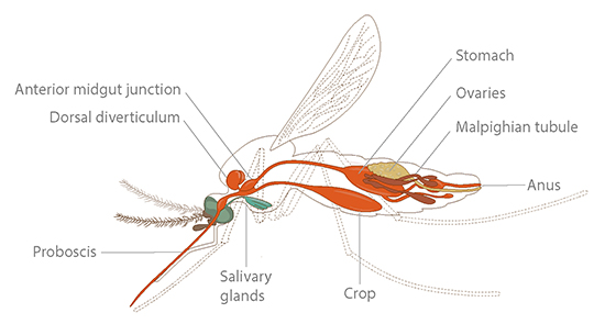
Labeled illustration of the internal anatomy of a female mosquito. Illustration by Karolina Mikołajczyk.
Looking at the Inside of an Adult Female Mosquito
| Proboscis | Tube-like mouth part used to suck up fluids. In females, this is strong enough that it can pierce skin. | ||
| Salivary glands |
Glands that make saliva. When saliva enters a mosquito bite, it helps make blood flow from the host, it numbs the skin, and it may pass along parasites, viruses, or bacteria that cause disease. |
||
| Dorsal diverticulum | A pouch in the anterior portion of the digestive system that is involved in sugar digestion. | ||
| Anterior midgut junction | A part of the anterior digestive system that helps meals to be processed in the correct part of the system. | ||
| Crop | A sac that stores sugary meals like nectar. | ||
| Stomach | A part of the posterior digestive system. Blood meals are sent directly to the stomach to be digested. | ||
| Ovaries | A part of the reproductive system, where eggs develop and are stored. | ||
| Malpighian tubule | An organ involved in the regulation of body water and hydration, as well as in getting rid of waste. | ||
| Anus | The end of the digestive tract, where wastes are removed from the body. | ||
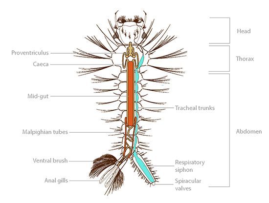
Labeled illustration of the internal anatomy of a mosquito larva. Illustration by Karolina Mikołajczyk.
Looking at the Inside of a Larval Mosquito
| Head | Location of the eyes and brain, and where the antennae attach. | ||
| Thorax |
Midsection where the (6) legs and (2) wings will later attach. |
||
| Proventriculus | A muscular pouch that helps grind food before it reaches the midgut. | ||
| Caeca | Small projections in the digestive system that increase surface area to release special proteins or to absorb nutrients or water. | ||
| Abdomen | The hind part of the mosquito, where most of digestion, eliminating waste, and reproduction occurs. | ||
| Mid gut | The first site of digestion, the midgut makes most of the enzymes used in the digestive process. | ||
| Tracheal trunks | Organs used in breathing. In adults, these usually connect to the outside through spiracles. In aquatic larvae, they connect to the siphon. | ||
| Malpighian tubule | An organ involved in the regulation of body water and hydration, as well as getting rid of waste. | ||
| Ventral brush | A collection of hairs at the rear of the abdomen. Hairs are used for sensing. | ||
| Anal gills | Special organs that absorb water and that are also involved in absorbing oxygen sometimes. | ||
| Respiratory siphon | A breathing tube that larval mosquitoes use; it uses water tension at the surface to attach there. | ||
| Spiracular valves | Valves that can close off the end of the siphon, so it does not take on water. | ||
Read more about: All About Mosquitoes
Bibliographic details:
- Article: Mosquito Anatomy
- Author(s): Dr. Biology
- Publisher: Arizona State University School of Life Sciences Ask A Biologist
- Site name: ASU - Ask A Biologist
- Date published: 17 Jun, 2024
- Date accessed:
- Link: https://askabiologist.asu.edu/mosquito-anatomy
APA Style
Dr. Biology. (Mon, 06/17/2024 - 17:53). Mosquito Anatomy. ASU - Ask A Biologist. Retrieved from https://askabiologist.asu.edu/mosquito-anatomy
Chicago Manual of Style
Dr. Biology. "Mosquito Anatomy". ASU - Ask A Biologist. 17 Jun 2024. https://askabiologist.asu.edu/mosquito-anatomy
Dr. Biology. "Mosquito Anatomy". ASU - Ask A Biologist. 17 Jun 2024. ASU - Ask A Biologist, Web. https://askabiologist.asu.edu/mosquito-anatomy
MLA 2017 Style
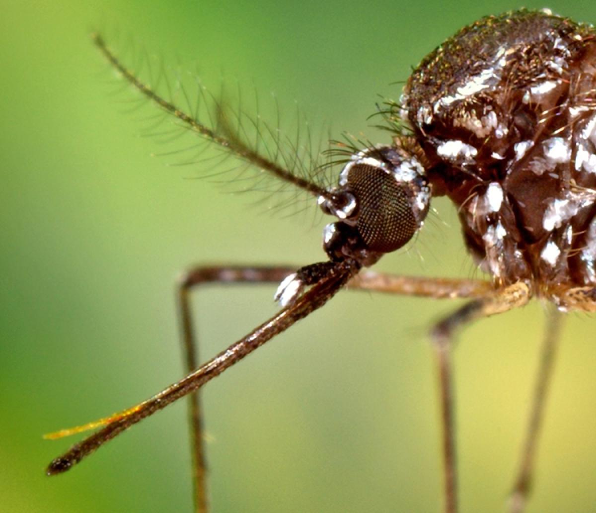
In this close-up image of a female Aedes aegypti, you can see different parts of the proboscis. The thin, needle-like section of the proboscis is the fascicle. The outer covering, which bends and pulls away from the needle when the needle is inserted, is called the labium.
Be Part of
Ask A Biologist
By volunteering, or simply sending us feedback on the site. Scientists, teachers, writers, illustrators, and translators are all important to the program. If you are interested in helping with the website we have a Volunteers page to get the process started.

