Illustrated by: Meredith Johnson
Tissue culture: Growing cells

Let’s try to remember the last time we were sick, shall we? Maybe it was a seasonal flu, or an episode of migraine, or a food allergy. Whatever the illness, there was most likely some medicine that came to our rescue. Fast forward a few days and most of us forgot that we had ever been sick. Our medicines indeed worked like a charm!
But how were these medicines created in the first place? To create a medicine, scientists first must understand how our cells act during an illness. Then, they must test out different chemicals that could change the behaviour of those ‘sick’ cells. But scientists would need living cells to perform these experiments. Where can one get such cells? This is where tissue culture comes into the picture.
Growing nerve cells

For a long time scientists have worked to understand the little universe that exists within our cells. Ross Harrison, who lived in the early 1900s, was one such scientist. He was mainly interested in nerve cells and how they grew in the early stages of embryonic development. But just like many other scientists in that era, he bumped into a big challenge - how would he study these cells?
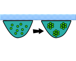
After many failed attempts to grow nerve cells in different kinds of solutions, he finally decided to get a bit creative. He tried to grow the cells in a drop of lymphatic fluid, suspended from an inverted glass - a method that came to be called ‘the hanging drop method’. The lymph that held all the essential nutrients acted as a nutritious broth for the growth of the cells.
When he viewed these cells under the microscope, he was able to study them without surrounding tissues blocking his view. Harrison’s experiments helped us learn the basics of how nerve cells develop. Perhaps more importantly, they opened the path for us to learn how to culture animal tissues.
What is tissue culture?

Tissue culture is a method of growing cells from plants and animals in the laboratory. Here we will specifically look at animal tissue culture. This means growing cells from animals including insects, mice, and human beings.
Tissue culture is one of the major tools used in biological research. This is because cells grown in tissue culture behave similarly to the organism they are taken from. Studying the cells can help us indirectly study the organism. But how do we grow or ‘culture’ cells? The cells in our body are part of an intricate framework. Oxygen and nutrients are supplied by the blood, and temperature and pH are controlled by complex processes. In order to grow our cells in the laboratory, we need to somehow recreate this complex system. Although this may seem difficult, scientists have been successfully doing this for many years now.

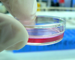
A scientist first extracts cells from the organism, for example from a patient biopsy sample. The cells are then placed in a tissue culture dish made of plastic or glass. The cells attach to the bottom of the dish and start dividing, spreading across the 2D surface of the dish. The growth of the cells depends on the following factors:
-
Nutrition: Cells need nutrients like sugars, amino acids, vitamins, and minerals to grow. Cells can get these when you grow them in nutritional media. The media could be a natural biological fluid or one that is made in the lab.
-
pH: The pH of the media is maintained by certain chemicals. The biomolecules inside the cell function best at the right pH (around 7.4).
-
Oxygen: The culture dish is placed in an incubator that can control the oxygen level in the air. This delivers a continuous supply of oxygen to the cells.
-
Temperature: Animal cells thrive best at 37℃, so the incubator maintains a temperature between 36℃ and 37℃.
-
Carbon dioxide: The incubator also maintains a level of around 5% carbon dioxide in the air. The carbon dioxide interacts with chemicals in the media and helps maintain the right pH.
How has tissue culture helped us?
Being able to isolate our cells and study them in the laboratory has helped us learn about cells and diseases. Without tissue culture, we wouldn't know as much about cancer, heart disease, and diabetes, for example. These diseases may have been a death sentence a few decades ago. Instead, we now have many medicines that can either cure these diseases or slow their progress.

Up until the 1900s, infections like polio and measles were famous for taking millions of lives. Today we are still at risk of viral infections like COVID and too many types of influenza or flu. Tissue culture has helped us better understand many viruses like these. It has also helped us make powerful vaccines against many of them.
If growing cells in 2D weren't innovative enough, we now have technologies where cells can be grown in 3D! This has given birth to the field of tissue engineering, where the aim is to make tissues artificially in the lab. These tissues can be used to treat injuries as well as diseases like diabetes.
Talking about innovation, 3D tissue culture has also made it possible to engineer products like meat, fish, and dairy foods. This is hoped to help reduce the environmental impact of these industries. Tissue culture has indeed opened our eyes to the bustling life inside our tiny cells and the immense possibilities that come with it.
Additional Images from Wikimedia Commons. Hamster cell petri dish image by the US NIH.
Read more about: Growing Cells
Bibliographic details:
- Article: Growing Cells
- Author(s): Sunaina Rao
- Publisher: Arizona State University School of Life Sciences Ask A Biologist
- Site name: ASU - Ask A Biologist
- Date published: 10 Aug, 2022
- Date accessed:
- Link: https://askabiologist.asu.edu/explore/tissue-culture
APA Style
Sunaina Rao. (Wed, 08/10/2022 - 20:54). Growing Cells. ASU - Ask A Biologist. Retrieved from https://askabiologist.asu.edu/explore/tissue-culture
Chicago Manual of Style
Sunaina Rao. "Growing Cells". ASU - Ask A Biologist. 10 Aug 2022. https://askabiologist.asu.edu/explore/tissue-culture
Sunaina Rao. "Growing Cells". ASU - Ask A Biologist. 10 Aug 2022. ASU - Ask A Biologist, Web. https://askabiologist.asu.edu/explore/tissue-culture
MLA 2017 Style

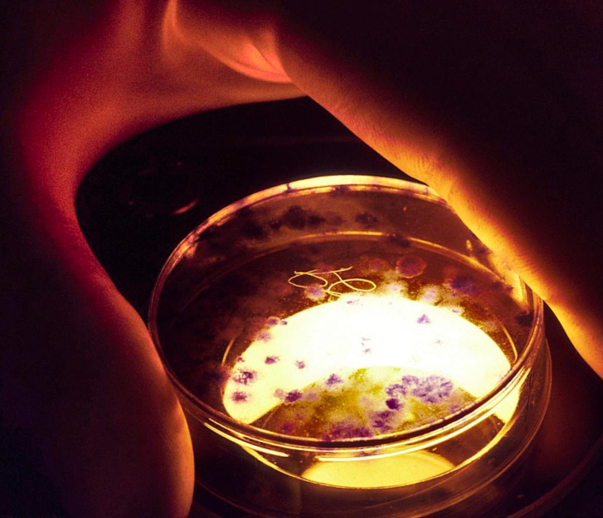
We use tissue cultures to understand how cells work. Here a scientist is studying hamster cells grown on a petri dish.
Be Part of
Ask A Biologist
By volunteering, or simply sending us feedback on the site. Scientists, teachers, writers, illustrators, and translators are all important to the program. If you are interested in helping with the website we have a Volunteers page to get the process started.
