Inner Space: The Final Frontier

[beeps – electronic lock and vault door opening]
Dr. Biology: This episode of Ask A Biologist is being pulled from our special collections that have been stored in our secret vault. This is Ask A Biologist, a program about the living world and I'm Dr. Biology. Allow me to start the show off with the opening lines from one of my favorite television shows. I've actually always wanted to do this, so, here's my chance.
[reverb effect voice]
Dr. Biology: Space ‑‑ the final frontier. These are the voyages of the starship Enterprise. Its continuing mission, to explore strange new worlds, to seek out new life and new civilizations, to boldly go where no one has gone before.
[end of reverb effect voice]
Dr. Biology: I'm sure many people know that these lines come from the show, which is the original Star Trek, or the more recent one. They've either seen the show, or there are a lot of movies out now. While the characters of Star Trek were exploring strange new worlds in outer space, there are also as many, if not more, strange new worlds right here on planet Earth.
To find them, you need only to look at the very small, or inner space, instead of outer space. In today's show, we'll be talking about inner space, microscopes and microscopists, or, as I like to say, micronauts, because they get to explore some of the most amazing worlds you can imagine.
With me are two micronauts. Doug Chandler, a professor in the ASU School of Life Sciences. Dr. Chandler is also the co‑director of the School of Life Sciences Bioimaging facility. You could think of it as the NASA for the School of Life Sciences. This is where the biologists at Arizona State University come to do their own exploring, finding their new life forms in worlds that are just unbelievable sometimes.
We also have a visiting scientist at ASU, Dr. Angela Goodacre, from Olympus. In case you didn't know it, Olympus makes more than cameras. They also build some amazing microscopes that are used by scientists around the world.
If you give us a moment, we'll also give you some great websites that you can visit and explore. Even if you don't have your own microscope, you'll be able to go and look at some of these worlds that biologists and scientists around the world are finding and posting up on the website.
Welcome to the show, Dr. Chandler.
Dr. Doug Chandler: Thank you, Dr. Biology.
Dr. Biology: And welcome, Dr. Goodacre.
Dr. Angela Goodacre: Thank you, Dr. Biology. I'm happy to be here.
Dr. Biology: Before we journey to the strange new world of inner space, let's talk just a bit about the microscope. How long have microscopes been in use?
Doug: Quite a long time. Originally, people didn't even have glasses or spectacles. An Italian invented the spectacle. Soon after, we all know about Galileo and his telescope. Although, there's actually another guy involved there that's not well known. Within a year later, in the 1600's, came a microscope. It wasn't until a hundred years after that, that van Leeuwenhoek got going in the Netherlands.
Dr. Biology: The microscope's been around for about 300 years. We know actually two characters in history that are really well known for the microscope. One is Robert Hooke, and the other one is Antonie van Leeuwenhoek. One was Dutch. Is that right, Doug?
Doug: Yes.
Dr. Biology: The other one was...
Angela: British.
Dr. Biology: Well, when we start with a microscope, Angela, what do we have to have to be a microscope?
Angela: Well, you need glass lenses that will allow you to blow up the image that you're looking at, so that you can see the fine detail. A long time ago, you had to be able to draw what you saw through this glass. Nowadays, of course, we use cameras. It's made life a lot easier for those of us who are artistically challenged.
Dr. Biology: That's a really good point. There weren't any cameras when they started. The first book that Robert Hooke produced was a wonderful series of illustrations based on what he saw through the microscope. One of them is from cork. I love the neat thing about this, because he could look at the little, what he called cells, in the cork, which actually became the name that we use today to talk about cells, the smallest unit of life.
The other really curious thing that I found, when I was looking up Robert Hooke, is he illustrated a really exquisite flea. Turns out, that flea was the one that was going through Europe at the time, and was responsible for the bubonic plague. In a way, it's good that he survived, considering he was actually illustrating fleas that could have been carrying the bubonic plague.
Let's talk at a little bit this other character, Antonie van Leeuwenhoek, the Dutchman. His microscope is a little bit different, though, than the compound microscope that we might have been talking about with Angela here.
Doug: Well, yes. He used a single lens which is almost like a drop of glass. He was a perfect craftsman in making these. In fact, he considered it a trade secret because he knew that he made the best glass lenses around at that time. No one else could equal seeing what he saw because of his lenses. He kept it a secret to himself. Because he made such good lenses, he saw all kinds of little animal life, animalcules as he called them. He loved the ones that moved.
Dr. Biology: Alright. What are some of the scientific breakthroughs that have happened because of the microscope? I mean we're talking 300 years. There have to be things that without the microscope, we would have been stuck.
Doug: Well, one of the biggest things is the organelles within the cell. Hooke may have been describing whole little animals, and van Leeuwenhoek little single cells. Soon the lenses became even better than van Leeuwenhoek's. Although it took another hundred fifty years that people were seeing things within cells, like the nucleophile chromosomes, mitochondria, and other such so‑called subcellular organelles.
Dr. Biology: Right. Organelles, meaning literally tiny organs. I think it's great, but I know there's some more out there. Angela?
Angela: Without the microscope, in vitro fertilization would not have been possible. Parents who couldn't have a baby by normal means, were able to have a baby conceived in a test tube, and then grow up in the mom.
Dr. Biology: We just recently had a big push on standing up against cancer, a really big telethon. It's something that we've been battling globally. The microscope has a really important role with the study and the possible cure, and/or treatment for cancer.
Angela: Yes it does. You can take cancer cells, and try out new forms of chemotherapy. These are chemicals that will selectively kill cancer cells, and leave normal cells unharmed. When doctors are trying to develop new therapies for cancer, sometimes they will do this with cells that are grown in a dish. Then, they will observe them in a microscope, and treat those cells with these chemicals, to see if these represent an effective cure for cancer.
Dr. Biology: Not only are there drugs that are being able to be designed to treat cancer, another problem with cancer, from what I understand, is finding out when someone has it. Not only finding out when someone has it, but finding out early on. I believe you actually were talking about some really interesting possibilities using a microscope.
Angela: Yes. There are ways to look inside the human body using modified microscopes. Probably some of your listeners will have parents or grandparents who go and get a colonoscopy. That can show very early signs of cancer at a time when it's early enough to treat that, so that your parent or grandparent will be cured.
That's a different type of microscope. It's used in the human body. The other thing that microscopes can be used for is to look at cells that are taken from somebody who has a cancer. The doctors can look at those cells in detail, discover what kind of cancer it is, and what's the best treatment for that cancer.
Dr. Biology: Right. When you mentioned about the microscopes inside the body it brought to mind the fact that a lot of us think about the microscopes we see in either high school when we're growing up or maybe when we went to college. They have a certain picture in their mind. These microscopes today have all sort of shapes. Obviously, if you're going to go inside the human body, they've got to be really small, and they have to be very flexible. That's pretty cool stuff.
Doug, you actually do quite a bit of research with cells yourself. You're a cellular biologist.
Doug: Yes. Even moving cells, like van Leeuwenhoek.
Dr. Biology: Can you talk just a little bit about what you've been doing in the lab?
Doug: We have some of the same interests as van Leeuwenhoek. That is, looking at how cells move.
Dr. Biology: So now are you taking a microscope, and maybe connecting a video camera?
Doug: Definitely.
Dr. Biology: As someone who came from the art background, it was curious to me as I was a photographer ‑‑ I am a photographer ‑‑what happened was, for me getting into microscopy was very easy because all I was doing is sticking another part of an instrument onto, basically, a camera.
Angela: A microscope is pretty much like one of those zoom lenses on your camera. You press a button, and you zoom in, and you can see things that are far away. With the microscope, we're not so much looking at things that are far away, but we use that zooming in capacity to make the image larger, so that we can see more detail in the image.
Dr. Biology: Right. Where we first see just the surface of skin, we zoom in a little bit further and we see the cells making up the skin. We zoom in a little bit further and we look inside the cells and we see the organelles that are inside there. We can go into one of the organelles.
For example, we can go into the nucleus and we can see the DNA that's inside there. As we go further and further in, we get more details.
Doug: That's an excellent analogy. The components of a camera work very similar to those of a microscope.
You have glass lenses that magnify things and focus things. You have light gathering capabilities. You're trying to gather the light from a landscape and focus it onto film. That's going to be your image that's recorded on film or on your digital chip in your camera.
Then you have a permanent image that's a representation of what you or the camera saw. The same thing happens in a microscope. You attach the camera to the microscope. You just have an additional set of lenses to make things look bigger. You focus them on the solid state chip or film and you've got an image that's preserved.
Dr. Biology: I see these wonderful images and I said we'd talk about some websites out there that people can go to and explore. One of them, Angela, is the company you work for. It's called Olympus Bioscapes.
I don't want people to miss the address. Don't worry about the "http" stuff. It's just olympusbioscapes.com. There are some amazing pictures up there. Now you said there are videos, too?
Angela: Yes. The Bioscapes Competition is organized by Olympus. Other microscope companies, like Nikon, also have competitions.
The one that is organized by Olympus, called Bioscapes, is looking for images that are taken using light microscopes. They are looking for scientific interest, but also the artistry behind it, whether it is an interesting composition.
This is open to all kinds of scientists throughout the world. You can submit either still images or you can submit movies.
Nowadays, with light microscopy, we like to be able to take pictures of living cells and even moving animals. Little animals and even big animals, anything that you take with a microscope you can submit for this competition.
Dr. Biology: All right. You mentioned Nikon. Nikon has what's called "The Small World" and that's nikonsmallworld.com. If you want to learn more about microscopes themselves, you want to go to a site called Molecular Expressions.
The best way to find that is type those two words in Google and you'll find the site. They have tutorials and they have movies and they have pictures and they have ways to use microscopes. It's a great place to go.
Also, Ask A Biologist has always had a gallery. We have a mystery image gallery. You can go in there and there are some really spectacular images. One of them, Doug, is work from your sea urchins. It's the one, it looks very out‑of‑this‑world. It looks like a bunch of planets.
Another one is ‑‑ well, I like to say that science is literally at our fingertips. This one is sweat on a fingertip. It's a scanning electron microscope picture of sweat on a fingertip. You've got to go see it.
Finally, we've added some really great galleries that allow you to now zoom in and zoom out as if you had your own private microscope. Even if you don't have one in school or if you don't have one at home, stop in to Ask A Biologist. You can start first with bird feathers and we're going to have one up on pollen grains. Go there and I know we're going to have a lot more in the future.
Doug: I'd also like to encourage you, if you find Ask a Biologist interesting and some of those pictures fascinating, you, too, can have your own microscope at home. There are microscopes available at virtually every store that caters to "playing with science".
I remember as a little kid I got one of these microscopes, probably from Sears or something like that. I was daring enough to prick my finger and look at my own blood underneath it.
Dr. Biology: Oh, really?
Doug: Yeah. All these cells were moving around. It was really cool. I could relate to van Leeuwenhoek.
Dr. Biology: If you don't want to prick your finger, you can go out there and get some really scummy pond water. That's another wonderful thing to look at because you can find some things in a drop of water that you couldn't believe.
Now, let's go a little bit further here with the world of microscopy. We began to talk about, they don't all look the same. A microscope is not just like every other microscope.
If you imagine walking into a hardware store and you look in the tool aisle, you might see a dozen different kinds of hammers and you might see at least that many kinds of saws. Even though they are all called hammers and they are all called saws, they each have different purposes. They work better for some situations than others. Let's talk about the different types of microscopes.
Angela, could you talk about the different basic ones that we have?
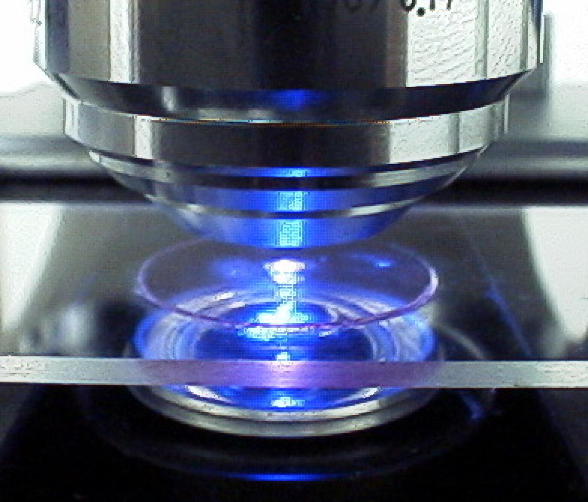
Angela: Many microscopes use light. There's light that is just white light and then you look at a pink or purplish colored dye on a piece of tissue. Then there are other microscopes that use a different kind of light. This is called fluorescence, just like fluorescent lights that you might have in your home.
But fluorescence can be made in many different colors. You can put chemicals into a cell, which will attach themselves to very specific things within the cell.
Dr. Biology: These are the little organelles.
Angela: These are the organelles or even specific molecules. You can tag these molecules or organelles with a color that represents that molecule or that organelle.
Then you can look using fluorescence microscopy and you can watch individual organelles move around the cell. You can see other cells interact with the cell that you're looking at. You can see cells in the bloodstream. You can see molecules moving between cells.
You know what you're looking at because every molecule of a certain type is going to be labeled or tagged with a specific color.
Dr. Biology: Oh, yes. It's kind of like if you got to look into the cells and you get to see a bunch of things [audio breakup] have red, some of them might be green, some are blue. If we're going to talk about an organelle, let's pick one.
For example, Doug, you work with sperm. Sperm have to do a lot of swimming, which means it takes a lot of energy. They have to have a lot of mitochondria.
We might have a particular kind of label, these kinds of chemicals that attach themselves to the mitochondria, so we can see how much mitochondria is in that particular sperm cell, right?
Doug: That's right. Sperm cells have a whole group of mitochondria just below what is called a flagellum. That's really just the tail that whips back and forth and makes the sperm swim. We could use a colored dye that's fluorescent and that's specifically designed to be taken up by mitochondria and color them red.
Angela: Then you can color the nucleus blue and you could color other parts of the cell yellow and green and you can look at the way how all these organelles interact with each other. One of the cool things about using cameras on a microscope now is that we can use colors that are invisible to the eye.
Just like insects can see into the ultraviolet range, there are colors that are invisible to us but they are perfectly visible to a bumblebee looking for a flower to pollinate.
We can extend our range of colors, our palette of colors, if you think like an artist palette, not only for the colors of the rainbow from red to violet, but we can go infrared and we can go ultraviolet. Some of our cameras can record these colors for us.
Dr. Biology: We have a color spectrum on the Ask A Biologist website. If anybody wants to see that spectrum including the ultraviolet and the infrared, they can do that. I recommend it because it gives you an idea of how much we would be missing if we couldn't do that.
We have light microscopes that can see the visible light that's visible to humans and we also have the microscopes that work with fluorescent colors. Those could be colors that we do see, and some of them we can't see but the cameras are able to see. What other kinds of microscopes do we have, Angela?
Angela: We have electron microscopes.
Dr. Biology: Electron microscopes?
Angela: Yeah. Instead of using light, which is made up of photons, the electron microscope looks at electrons.
Dr. Biology: OK. Doug, you're actually, if I'm not mistaking, one of the big areas you spend a lot of time in is electron microscopy. I think you've written a book on this, haven't you?
Doug: Yes. Both light and electron microscopy.
Dr. Biology: What's the title of it?
Doug: It's called "Bio‑imaging and Current Concepts in Light and Electron Microscopy".
Dr. Biology: Not everybody is going to go pick that up, but I'll tell you, it's got a lot of really cool pictures in it. Let's talk a little about light and electron microscopes. Why are we using electrons instead of these photons that come from the Sun?
Doug: It seems surprising because you can't even see electrons. The only way you can detect them is by film, you'd take a picture of electrons, or by a phosphorescent plate that will change electrons in the light.
Dr. Biology: Why electrons instead of photons? I can go get a light bulb and do that very easily. I'm assuming it's not as easy to make a microscope that uses electrons. Why would I want to use those?
Doug: Electrons seem like a particle to some scientists but they seem like waves to others. In the microscope we're always using waves. The electron, it turns out, the way it goes through our specimens and interacts to form an image of the specimen is acting in a sense similar to a photon.
There's one big, big difference, though. The wavelength of the electron is much smaller than that of the photon. That means it can see little details of the specimen that a photon could never see.
Dr. Biology: Right. Let me get this straight. You have photons that have these big waves and you have electrons that form these really tight waves and because of the wavelength they're able to see much more detail in really, really small things.
I bet someone's going to start mentioning a word that we use a lot but not necessarily the way we want to use it in the show, and that's resolution. What, on Earth, is resolution? This is what we're going to get to when we talk about the difference between looking at a light microscope and looking at an electron microscope.
Angela: Resolution is how close together two points can be, and yet you can still tell that they're two separate points. Higher resolution means a smaller distance between those two points.
Dr. Biology: If we're able to see something like two atoms as two separate atoms, that would be really good resolution.
Angela: It would be really good, and you wouldn't be able to do it with a light microscope.
Dr. Biology: What we typically look at with microscopes in school or anywhere else, what would be the best resolution we can possibly get just with a white light?
Doug: It would be a fraction of a micron, which seems small but you would have to put billions of atoms together to make that distance. Instead, we're looking at bigger things like subcellular organelles.
Dr. Biology: If electron microscopes are able to see much smaller detail than the light microscopes, why don't we just use electron microscopes? Why isn't that the only microscope we use?
Doug: It's hard to look at a live cell inside with the electron microscope because electrons only work in a vacuum.
Dr. Biology: Like out in space?
Doug: Yeah. Out in space.
Dr. Biology: What is it that we can see with an electron microscope that we can't see with a light microscope? Do you have some examples for me?
Angela: In the light microscope we can see an individual cell, we can see the nucleus, we can see the mitochondria. But in order to see the cell membrane itself we'd have to turn to electron microscopy. Another example would be, we can see bacteria in the light microscope but we can't see viruses. That's why we have to go to electron microscopy.
Doug: Microscopy, especially electron microscopy, is important to the medical sciences. At the light level you look at the tissue that might have a pathology, maybe cancer. You recognize that. Then you go to the electron microscopy level and find out what parts of the cell are unusual.
For example, there are whole new sets of cellular structures that have been seen only with the electron microscope. The cell membrane, that covering that surrounds the cell that's so important in the cell not only interacting with its neighbors but it's the part of the cell that is attacked by viruses and bacteria, it wasn't even seen until electron microscopy was invented.
It would be like, we know what your house looks like and we know it has a kitchen and a dining room and a living room, and we know there's a really gigantic big screen TV in the living room but we don't even know what kind of walls make up your house. We couldn't see them. They're so thin that our microscope can't see them. With the electron microscope, we can tell that you must live in a brick house.
Dr. Biology: OK, you two micronauts, before we get out of here, I always ask three questions. I'll start with Angela. The first one is, when did you first know you wanted to be a biologist or a scientist? Was there a particular spark in your life that you can remember?
Angela: I think that I became a scientist because I always liked math.
Dr. Biology: Really?
Angela: Maybe, I was being a little bit lazy. I could sit down and take a math exam without having done any studying, whereas for history I had to hit the books and really study hard.
Dr. Biology: So your introduction into science was through mathematics?
Angela: It was, yes. That started just by adding up numbers. I have always been fascinated by numbers. Then, when I started looking through the microscope, it just seemed to make sense. It's the sense of discovery.
Dr. Biology: How about you Doug? What was the spark for you?
Doug: It just shows that people who end up in science can get there by many routes. I had two favorite hobbies since I was in high school. One, I really liked art, and I still do some painting and things like that.
Two, I really like biology and I was influenced so favorably by one of my favorite high school teachers who taught Advanced Biology. Probably all of you know, you have a few favorite teachers yourself, and you know how much they impart to you in excitement.
That's the kind of excitement I had when I met my high school biology teacher. It seemed inevitable that somewhere in the distant future that I would bring art and biology together and become a microscopist.
Dr. Biology: I can see you are both hooked on this, and you're passionate about it. My next question is what are you going to do when I take it all away from you? You can't be a biologist or a scientist. Certainly, you can't use a microscope. What are you going to do, what are you going to be, Angela?
Angela: I'm going to be an explorer.
Dr. Biology: An explorer?
Angela: Yeah, go all over the world looking for wildlife, plants, animals.
Dr. Biology: OK. I'm going to let you go away with that because I think there are some biologists that get to do that, too. I think you brought up a really good point. A lot of people if they want to do some exploring, they want to travel, biology is a great way to do it.
All right, Doug. I might know the answer to this but I'm going to take it all away, no science, no biology, none of those things. What are you going to be?
Doug: I only get one choice?
Dr. Biology: You only get one choice.
Doug: Oh man, that's tough. Mine is very ordinary. It used to be that every kid wanted to be a policeman. I think most kids these days want to be a rockstar and that's what I want to be.
Dr. Biology: You want to be a rock star? I thought you were going to say you wanted to be an artist. I forgot to mention, Doug also has the cover of one of the Sols magazines. It's one of his paintings. It's a fabulous painting. You go into the Sols website, sols.asu.edu, and you can see some of these really cool images.
All right. One more question for the two of you. What advice would you have for someone who wants to become a biologist or just a scientist in general? Angela, even a mathematician?
Angela: To get very familiar with computers because every time we take an image through the microscope these days, it's going to be stored on a computer. You really need to have a good understanding of where your images are stored on a computer, how to get them, how to label them so you know exactly what you did to get that particular image. I think these are important things in preparing to be a scientist, particularly if you want to be a microscopist.
Dr. Biology: OK, and Doug?
Doug: My advice is more general. I think anything that you really want to do, you want to do because of motivation. Where do you get that motivation? I think you look to other people that are doing the things that you find interesting and that you would like to do.
If you think science is kind of neat, look around the web, then go to that science teacher that you think is maybe doing some really weird stuff or cool stuff, as the case may be. If you like art, find somebody who can tutor you in art, who really loves art. Whatever you like to do, go to a person that can make it exciting for you, who can give you the motivation to really work on it.
Dr. Biology: Professor Chandler, thank you for visiting us.
Doug: Thank you.
Dr. Biology: Dr. Goodacre, thank you for joining us on Ask a Biologist.
Angela: Thank you.
Dr. Biology: You have been listening to Ask a Biologist, and my guests have been Professor Doug Chandler from the ASU School of Life Sciences, and our special guest Dr. Angela Goodacre from Olympus.
The "Ask A Biologist" podcast is produced on the campus of Arizona State University and is recorded in the Grassroots Studio housed in the School of Life Sciences, which is an academic unit of the College of Liberal Arts and Sciences.
And remember, even though our program is not broadcast live, you can send us your questions about biology using our companion website. The address is askabiologist.asu.edu or you can just Google the words "Ask A Biologist". I'm Dr. Biology.
Bibliographic details:
- Article: Inner Space: The Final Frontier
- Author(s): Dr. Biology
- Publisher: Arizona State University School of Life Sciences Ask A Biologist
- Site name: ASU - Ask A Biologist
- Date published: 17 Oct, 2015
- Date accessed:
- Link: https://askabiologist.asu.edu/podcasts/inner-space-final-frontier
APA Style
Dr. Biology. (Sat, 10/17/2015 - 16:17). Inner Space: The Final Frontier. ASU - Ask A Biologist. Retrieved from https://askabiologist.asu.edu/podcasts/inner-space-final-frontier
Chicago Manual of Style
Dr. Biology. "Inner Space: The Final Frontier". ASU - Ask A Biologist. 17 Oct 2015. https://askabiologist.asu.edu/podcasts/inner-space-final-frontier
Dr. Biology. "Inner Space: The Final Frontier". ASU - Ask A Biologist. 17 Oct 2015. ASU - Ask A Biologist, Web. https://askabiologist.asu.edu/podcasts/inner-space-final-frontier
MLA 2017 Style

Explore inner space yourself with our Zoom Gallery.
Be Part of
Ask A Biologist
By volunteering, or simply sending us feedback on the site. Scientists, teachers, writers, illustrators, and translators are all important to the program. If you are interested in helping with the website we have a Volunteers page to get the process started.
